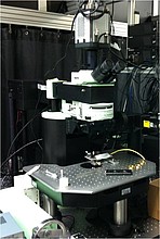
In vivo imaging/two-photon microscopy
In vivo imaging enables non-invasive examination of living organisms and helps to understand metabolic processes, immune responses and disease related changes in the body.
Two-photon microscopy is the approach to study structures and functions of living cells and tissues up to a very higth depth using fluorescent dyes for visualization or autofluorescence signals. Due to the multiphoton absorption, the background signals are strongly suppressed and the photo toxicity is strongly reduced.
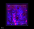
Vessels (red) are visualized by Dextran coupled to the dye TRITC in the skin, whereas the blue autofluorescence signal demarcates collagen bundles of the dermis.
